Neuroimaging technologies
We develop cutting-edge non-invasive and invasive brain imaging and neuromodulation methods.
Magnetic resonance imaging (MRI)
MRI has millimeter spatial resolution and versatile contrasts. MRI has been extensively used in clinical medicine and neuroscience studies to provide structural, functional, and metabolic information. We are devoted to advancing MRI technology in order to improve its spatiotemporal resolution and sensitivity. We aim to develop a tailored combination of receiver coil array, spatial encoding magnetic fields, main magnetic field, pulse sequences, and image reconstruction algorithms in order to optimize MRI in different neuroscience and clinical applications.
Electroencephalography (EEG), magnetoencephalography (MEG), and stereo-EEG (SEEG)
EEG and MEG are methods of studying neural activities non-invasively using extracranial measurements of magnetic fields and electric potentials, respectively. We continuously develop EEG and MEEG methods in order to better understand how the human brain works via studies of MEG/EEG source localization, neuronal oscillations, and multimodal (EEG/MEG/MRI) integration. We also developed methods of analyzing SEEG, an invasive recording of multiple electrodes implanted in medically refractory patients. These data allow for unprecedented spatiotemporal resolution of neuronal activities.
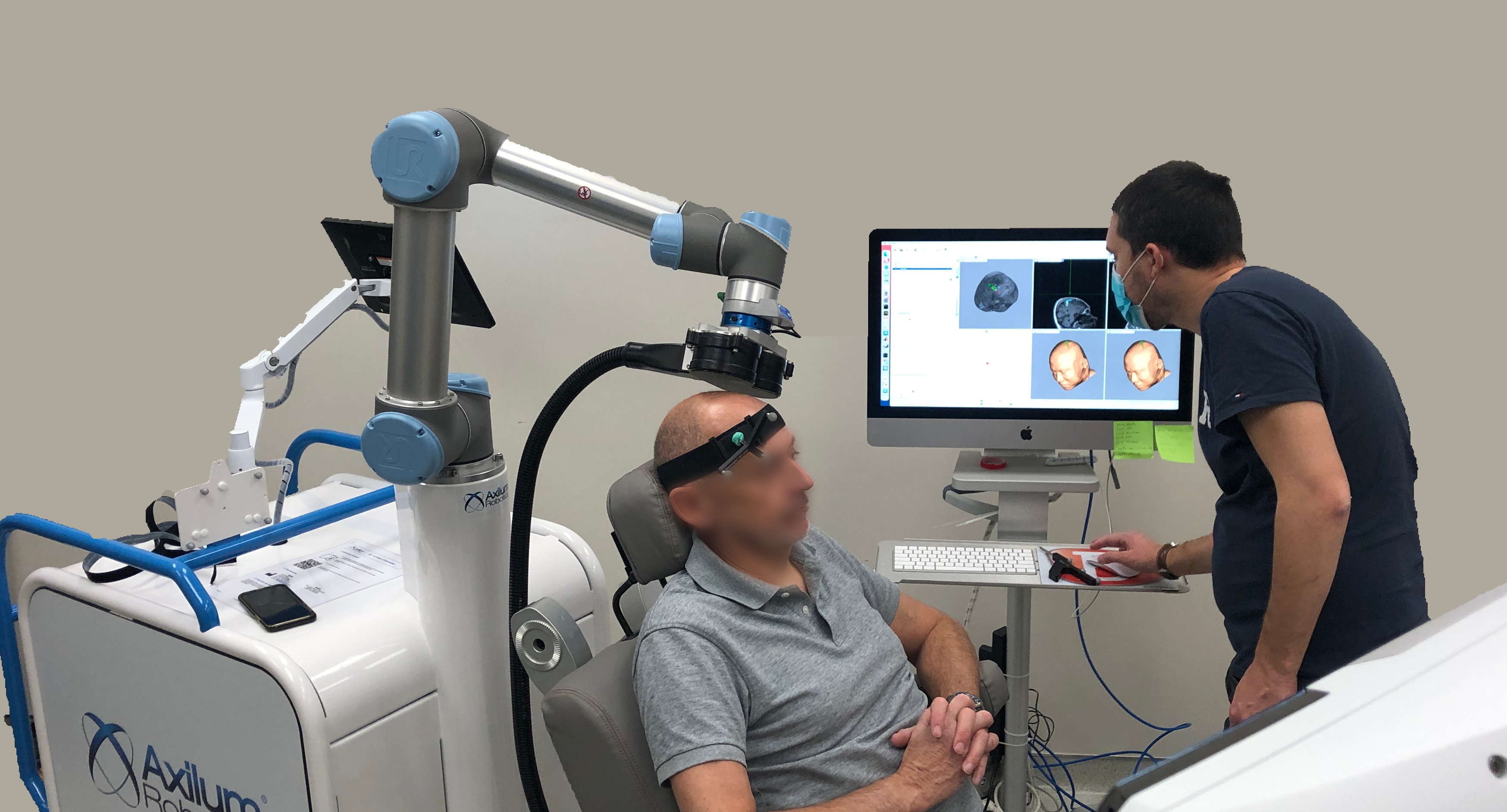
Transcranial magnetic stimulation (TMS)
TMS stimulates neural populations at the cortex via transient and strong magnetic pulses delivered by coils outside the head. We use neuroimaging (MRI/EEG/MEG) and navigation systems to excite and inhibit neural activity in a spatiotemporally accurate way.
Computational neural models
With rich datasets of brain activities characterized by neuronal, hemodynamic, and metabolic measures, we develop computational tools to reveal correlations and causal modulations between the brain and behaviors. Specifically, we aim to use advanced machine learning methods to develop models by combining large neuroimaging data sets and project-specific data sets with a smaller sample size.